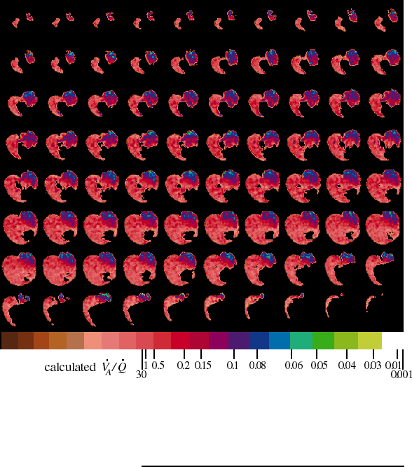
Image of obstructed ventilation is the quotient of two NMR images of SF6
in rat lungs. The left bronchus is partially obstructed. An obstruction-detecting image,
made from 1.5 hours of data collection while the rat breathed 25% SF6 : 75% O2, is divided
by a reference image, made from 1 hour of data collection while the rat breathed 80% SF6 :
20% O2. From top left to bottom right, eighty successive x-y planes, each with 72 by 74
pixels, are displayed from anterior (forward) to posterior (rear). The first row contains
the anterior tips of the lungs while lower down, the dark heart and mediastinum separate
the pink right lung from the purple and blue left lung. The large dark area in the lowest
planes is the diaphragm and liver. Pixel values range from 0 (black) to 1 (white), and are
divided into 21 color bands. The width of the color bands is one standard deviation for
individual pixels of that color. This measurement error is greater for higher pixel values
so the right color bands are wider than the left ones. The ventilation-to-perfusion ratio
(VA/Q) scale shows which colors represent different ratios. Like many lab rats, this one
has a small lobe of right lung in the posterior left thorax, which accounts for the pink
portion on the left side of the rat (right side of image).